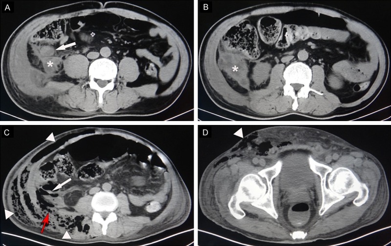
Figure 1.

Selected axial views of the abdominal computed tomography scan. A. A swollen appendix (white arrow) with abscess formation in the retroperitoneum (asterisk) was identified on first admission to local hospital. Notably, the right side abdominal muscles and psoas muscle were also swollen. B. The appendiceal abscess (asterisk) was approximately 5×5 cm in size. C. A second abdominal computed tomography showed a perforated appendix (white arrow) and gas and fluid collection extending from his retroperitoneal cavity to the subcutaneous layer of his right loin (white triangle). Notably, the inflammation passed through the inferior lumbar triangle (red arrow) to the flank and the lumbar area. D. Gas and fluid collection were also observed at the right lower limb (white triangle).
