Fig. 1.
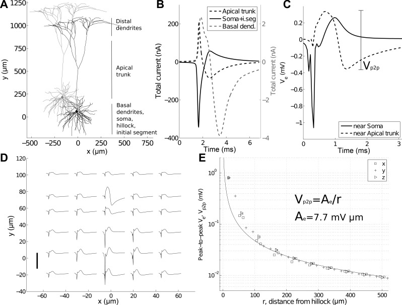
Characterizing electric potentials around a spiking neuron. A: multicompartment, 3-dimensional, layer V pyramidal cell model (Mainen and Sejnowski 1996). Duplicate model cell is shown in gray. B: summed membrane current for all compartments of the apical trunk, soma [including the hillock and initial axon segment (i.seg)], and the basal dendrites. C: 2 characteristic extracellular potentials (Ve). One is calculated at a distance 10 μm from the hillock (solid trace) and the other, ∼80 μm from the soma near the apical trunk (dashed trace). The peak-to-peak Ve close to the apical trunk is highlighted. D: Ve for 30 locations in the x, y plane of the neuron in A. Each trace is 3 ms in duration, and the scale bar is 1 mV. Traces are plotted at the spatial locations at which they were calculated. The soma is centered at (0, 10). E: peak-to-peak amplitude of Ve as a function of distance from the hillock. Locations were sampled along the x-, y-, and z-axes at regular intervals. The 1/r best-fit line (least-squares) and equation are shown.

