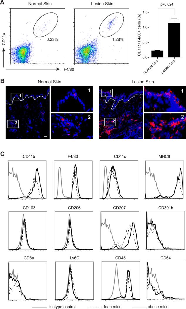
Figure 2. HFD promotes the accumulation of CD11c+ macrophages in obese mice.
(A) Analysis of the accumulation of CD11c+ macrophages in skin tissues from LFD-fed normal mice and HFD-fed lesion mice by flow cytometry. Average percentage of CD11c+ macrophages in skin is shown in the right panel.
(B) Frozen sections of skin tissues from LFD-fed normal mice and HFD-fed lesion mice were stained with F4/80 mAb (red) and DAPI (blue) for confocal microscopy analysis. Magnified fields of selected areas are shown in the right panel. Scale bars represent 10μM.
(C) Phenotypic characterization of HFD-elevated skin macrophages by flow cytometric analysis.
Data are shown as mean ± SEM and representative of at least three experiments. See also Figure S1.

