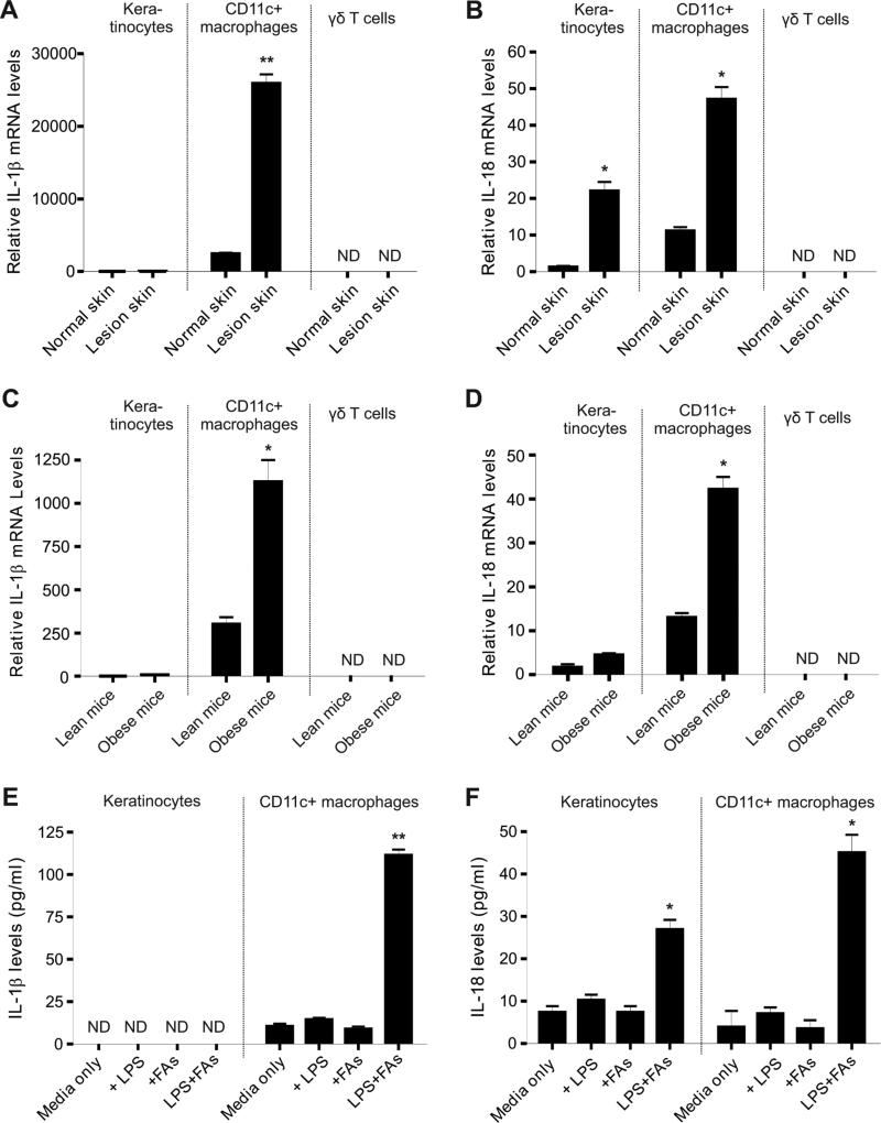
Figure 3. Upregulation of IL-1β and IL-18 production in HFD-induced skin lesions.
(A-B) Keratinocytes, CD11c+ macrophages and γδ T cells were separated by a flow sorter from either normal skin tissues or HFD-induced lesional skin tissues. Relative levels of IL-1β mRNA (A) and IL-18 mRNA (B) were determined by quantitative real-time PCR (*, p<0.05; **, p<0.01).
(C-D) Keratinocytes, CD11c+ macrophages and γδ T cells were separated by a flow sorter from feltwheel-induced lesional skin tissues from lean or obese mice. Relative levels of IL-1β mRNA (A) and IL-18 mRNA (B) were determined by quantitative real-time PCR (*, p<0.05).
(E-F) Measurement of IL-1β (E) and IL-18 (F) by ELISA in supernatants of primary keratinocytes or CD11c+ macrophages stimulated with designated conditions for 24 hours (LPS, 100ng/ml, FAs, 200μM palmitate) (*, p<0.05; **, p<0.01).
Data are shown as mean ± SEM and representative of at least three experiments. See also Table S1 and Figure S2.

