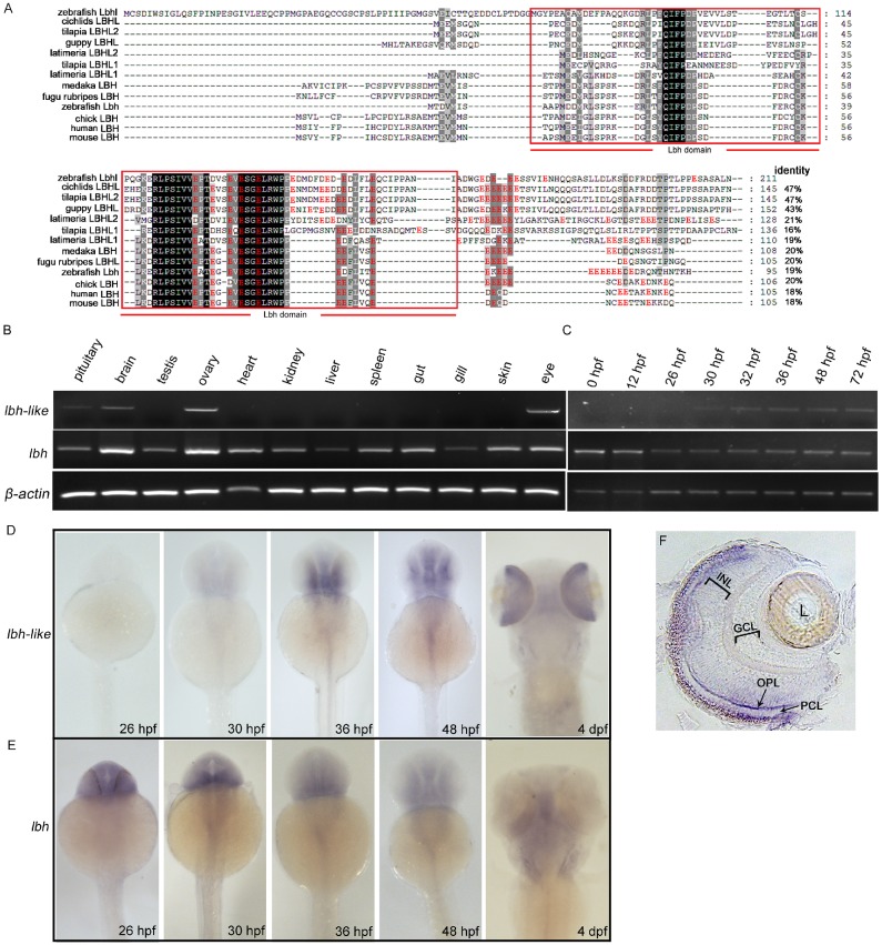
Figure 1.
Molecular and expressional characterization of lbh-like (lbhl) and lbh in zebrafish. (A) Multiple amino acid sequence alignment of zebrafish Lbh-like (Lbhl) and Lbh as well as other vertebrate LBHL and LBH. The black and dark shadings indicate identical sequences. The C-terminal glutamate-rich (red color) conserved LBH domain is labeled by red box. The GenBank IDs for these proteins used in this study are as follows: zebrafish (Danio rerio) Lbhl, XP_001336471; cichlids (Haplochromis burtoni) LBHL, XP_005927379; tilapia (Oreochromis niloticus) LBHL2, XP_003441738; guppy (Poecilia Formosa) LBHL, XP_007575999; latimeria (Latimeria chalumnae) LBHL2, XP_006000505; tilapia (Oreochromis niloticus) LBHL1, XP_005464744; latimeria (Latimeria chalumnae) LBHL1, XP_005988936; medaka (Oryzias latipes) LBH, XP_004082531; fugu rubripes (Takifugu rubripes) LBHL, XP_003962799; zebrafish (Danio rerio) Lbh, NP_956814; chick (Gallus gallus) LBH, NP_001026209; human (Homo sapiens) LBH, NP_112177; mouse (Mus musculus) LBH, NP_084275. (B) Semi-quantitative RT-PCR detection of lbh-like and lbh expression in adult tissues. (C) Semi-quantitative RT-PCR detection of lbh-like and lbh expression during embryogenesis. The β-actin was used as internal control. (D, E) Whole-mount in situ hybridization with lbh-like antisense probe (D) or lbh antisense probe (E). (F) Cryosection of eye from embryo hybridized with lbh-like antisense probe at 4 dpf stage. INL, inner nuclear layer; GCL, ganglion cell layer; OPL, outer plexiform layer; PCL, photoreceptor cell layer; L, lens. Dorsal views.

