
Figure 2.
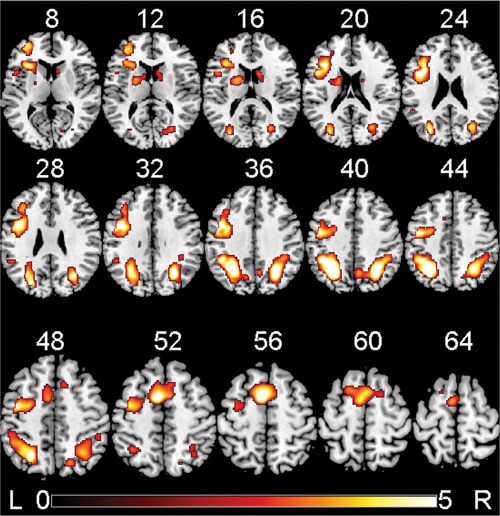
False discovery rate corrected significant clusters for the main effect of the delayed minus immediate memory conditions pooled across three digits, five digits, and seven digits per stimulus and across both groups combined, detected by the univariate SPM8 second‐level GLM analysis. Brain activations are overlaid in color on axial slices of the MNI template brain. The number above each slice indicates slice location (mm) of the MNI z coordinate. Scale on color bar represents voxel t values. The viewer's left (L) side of each slice is left hemisphere of the brain, and right (R) side of each slice is right hemisphere of brain.