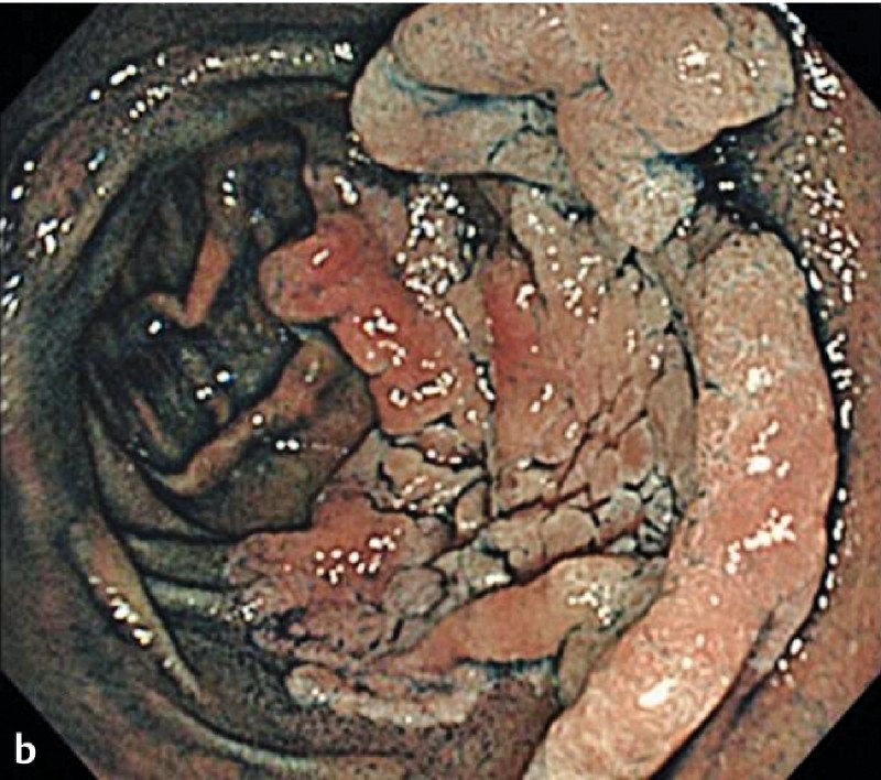
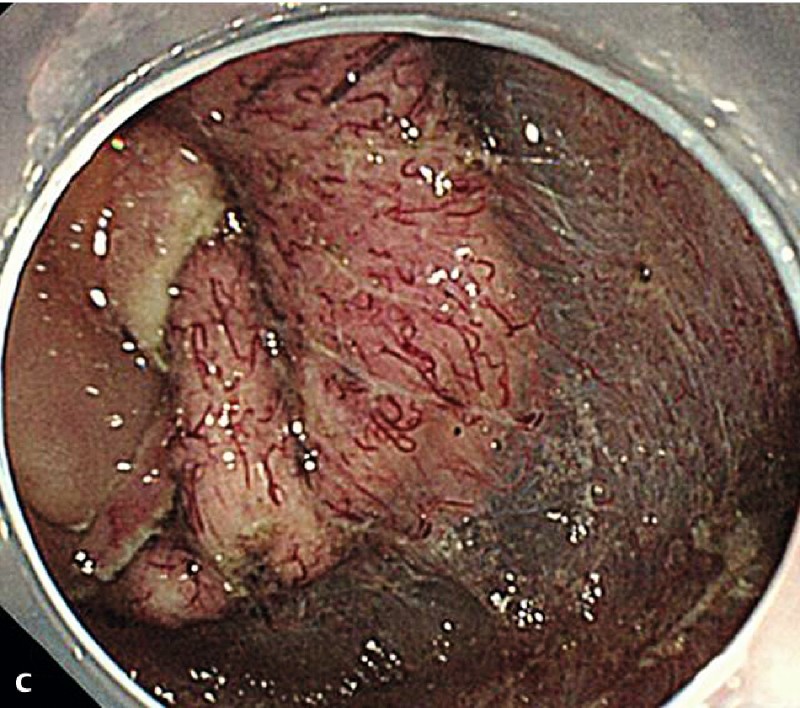
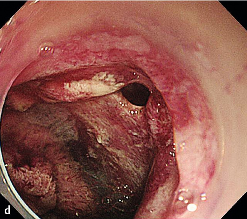
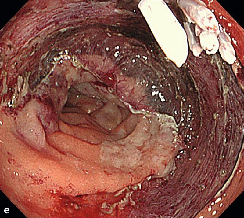
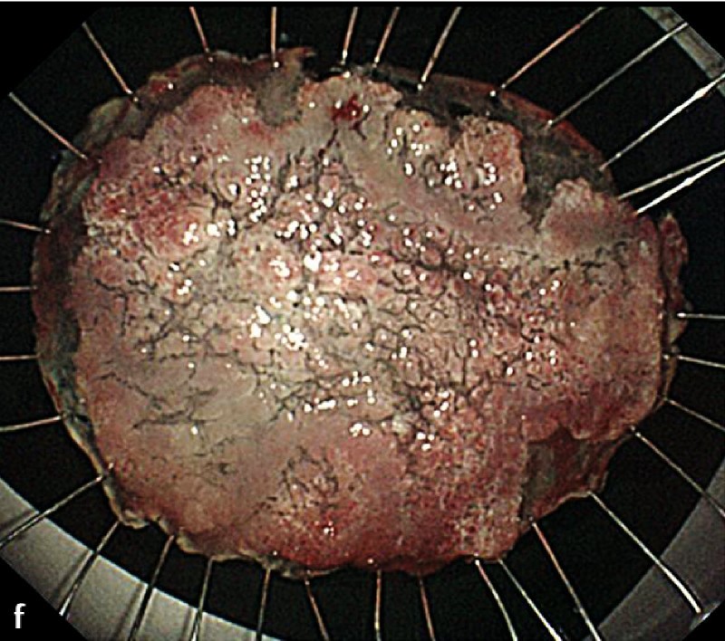
Fig. 2 A representative case of duodenal endoscopic submucosal dissection (ESD). a A large superficial neoplasm occupying two-thirds of the descending portion of the duodenum. b Chromoendoscopy finding after indigo carmine dye spraying. c Abundant blood vessels were observed in the submucosal layer. d A perforation occurred during the ESD procedure. e The perforation was closed by endoscopic clipping and successfully managed conservatively. f Resected specimen: 57 × 44 mm, well-differentiated adenocarcinoma with adenomatous component, depth intramucosal, no lymphatic invasion (ly0), no vascular invasion (v0), margin-negative.
