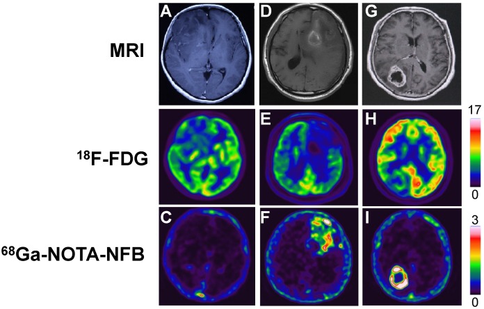
Fig 3.
Comparison of MRI, 18F-FDG PET and 68Ga-NOTA-NFB PET in three different glioma patients. In the upper row, A & G are enhanced MRI by administration of gadolinium contrast agent and D is MRI without contrast enhancement. The middle row shows 18F-FDG PET and the lower row shows 68Ga-NOTA-NFB PET. The first column (A, B & C): Patient No. 2, F, 30 y, grade II; the second column (D, E & F): Patient No. 3, M, 61 y, grade III; the third column (G, H & I): Patient No. 4, F, 60 y, grade IV.

