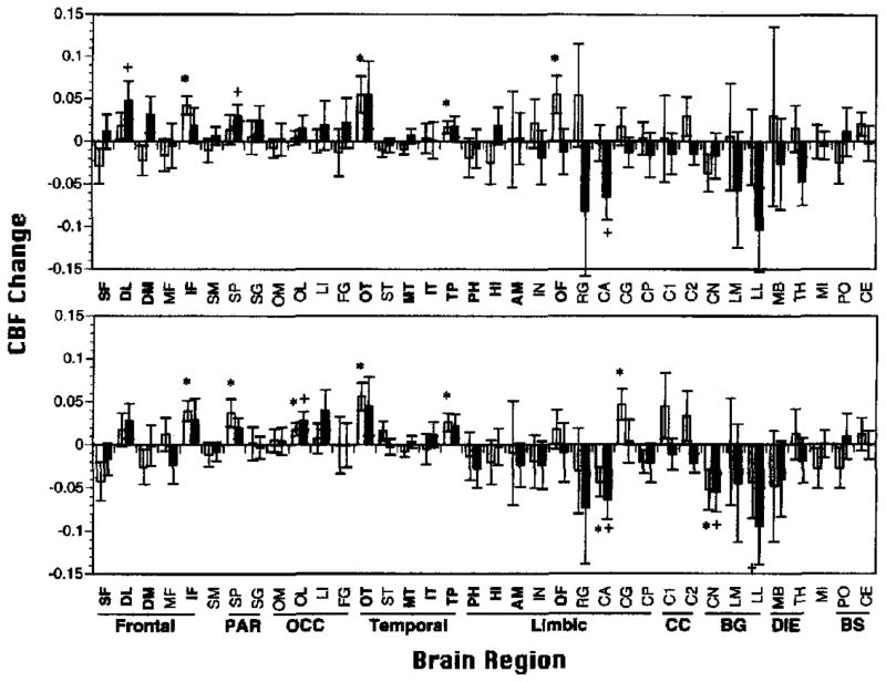
Figure 6.
Mean (±SEM) relative regional cerebral blood flow change (rCBFΔ; baseline subtracted) for all 36 regions of interest (ROIs) for Paired Associate Recognition Test (PART; top graph) and Wisconsin Card Sorting Test (WCST; bottom graph) for 15 demographically matched controls (open bars) and 15 patients with schizophrenia (shaded bars). SF = superior frontal; DL = dorsolateral prefrontal; DM = dorsomedial prefrontal; MF = mid-frontal; IF = inferior frontal; SM = sensorimotor; SP = superior parietal; SG = supramarginal gyrus; OM = occipital-medial; OL = occipital-lateral; LI = lingual gyrus; FG = fusiform gyrus; OT = occipitotemporal; ST = superior temporal; MT = mid-temporal; IT = inferior temporal; TP = temporal pole; PH = parahippocampal gyrus; HI = hippocampus; AM = amygdala; IN = insula; OF = orbital frontal; RG = rectal gyrus; CA = cingulate gyrus-anterior; CG = cingulate gyrus; CP = cingulate gyrus-posterior; Cl = corpus callosum-anterior; C2 = corpus callosum-posterior; CN = caudate nucleus; LM = lenticular-medial (globus pallidus); LL = lenticula-lateral (putamen); MB = mammillary body; TH = thalamus; MI = midbrain; PO = pons; CE = cerebellum; PAR = parietal; OCC = occipital; CC = corpus callosum; BG = basal ganglia; DIE = diencephalon; BS = brainstem. Regions that were analyzed in the primary analysis are in bold. *p < .05 controls; +p < .05 patients; all two-tailed.

