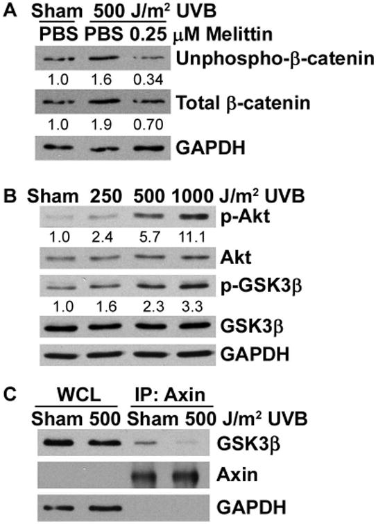
Figure 5. UVB disrupts the β-catenin phosphorylation/destruction complex.

(A) 308 cells were pre-treated with 0.25 μM melittin for 1 hr and then sham irradiated or exposed to 500 J/m2 UVB for 8 hrs. (B) 308 cells were sham irradiated or exposed to 250, 500, or 1000 J/m2 UVB and incubated for 8 hrs. Cell lysates were subjected to immunoblot analysis with antibodies specific for the indicated proteins. Representative blots are shown and numbers indicate fold change of proteins normalized to GAPDH. (C) 308 cells were sham irradiated or exposed to 500 J/m2 UVB for 8 hrs. Cell lysates were immunoprecipitated with an antibody against Axin and probed for GSK3β by Western blot. WCL, whole cell lysate.
