Figure 3.
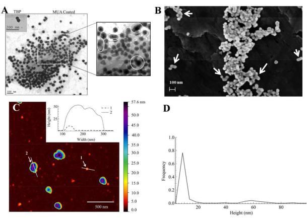
Electron and atomic force microscopy characterization of TBP coated GNPs produced using 50 pM citrated ~ 23 nm seeds in aqueous precursor solutions containing L31(8 mM)/F68(4 mM) and ≈ 1 mM Au(III) after 7 days incubation at room temperature. (A) Transmission EM of TBP coated (inset) and 11-mercaptoundecanoic acid (MUA) coated GNPs. (B) Scanning EM image of TBP coated GNPs, where the white arrows show the compact aggregates. (C) Representative AFM (512 × 512 pixels) 2 × 2 μm2 image of TBP coated GNPs. (D) Particle height distribution (N = 401), from AFM analyses, with two main population means at (9.3 ± 1.9) nm and (59 ± 8) nm, respectively.

