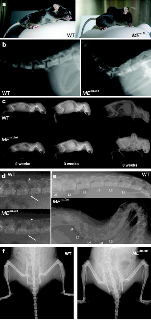
Figure 2. Severe spine degenerative changes associated with kyphoscoliosis in MEm1/m1 mice.
a) Photographs of WT and MEm1/m1 littermates, showing lordosis and kyphosis in MEm1/m1; note also dorsiflexed tail in MEm1/m1. b) Lateral radiographic view of adult WT and MEm1/m1 littermates, displaying junction between sacral and caudal vertebrae. c) Lateral radiographic views of WT and MEm1/m1 littermates at 2, 3, and 8 weeks, as indicated. d) Higher magnification at 2-weeks: note narrowing of joint spaces between L4 and L5, arrow (anterior) and arrowhead (posterior). e) Higher magnification of lateral view of lumbar spine at 10 weeks. f) Anterior-posterior (AP) view at 10 weeks. Note in MEm1/m1 mouse, the marked scoliosis and the loss of distinct margins (spontaneous fusion) between vertebrae throughout the lumbar, sacral, and caudal regions.

