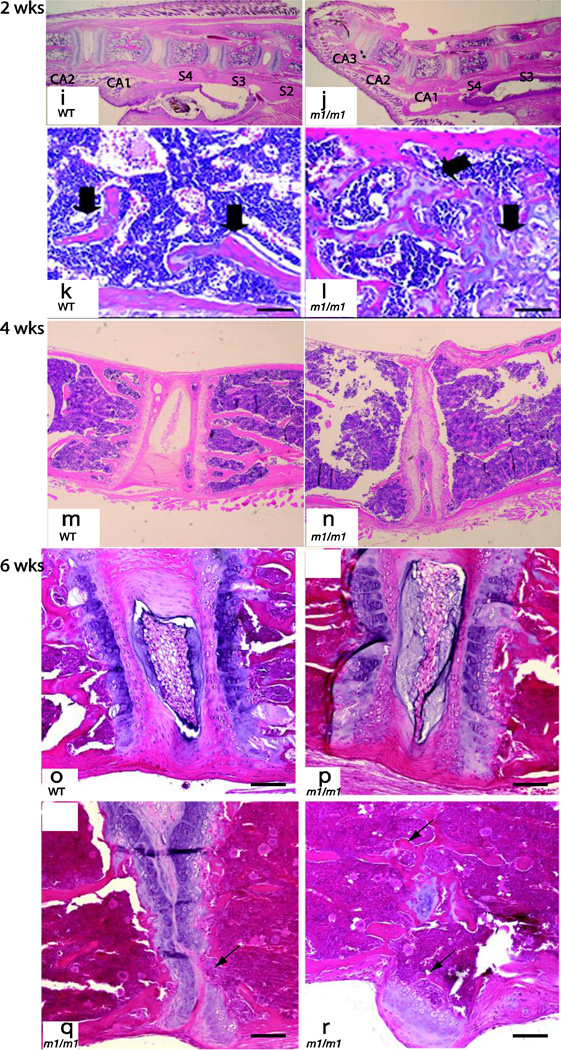
Figure 4. Histologic analysis of spine in MEm1/m1 mice.
Representative H&E-stained sections from one day (a–d), 10 day (e–h), two (i–l), four (m and n), and 6 week-old (o–r) mice show a range of spinal abnormalities in mutants. Panels a–d show sacral region at low and high power, with no demonstrable difference between WT and MEm1/m1 mice at one day of age. At 10 days (panels e–h), there is evident osteopenia in the lumbar region of MEm1/m1 mice, though differences are subtle. At 2 weeks, marked delay of endochondral ossification of lumbar vertebral growth plates is present in the mutant (l, black arrow). In the wildtype littermate, cartilage is restricted to physeal plates (k). At 4 weeks, lumbar discs show partial collapse in MEm1/m1 mice (panel n). Panels o–r illustrate various disc changes in the lumbar spine at six weeks. Compared to WT, MEm1/m1 mice show a progression from relatively normal disc morphology (p), to narrowed intervertebral space, to disappearance of the nucleus pulposus and loss of cartilage (arrow, q), and fusion of vertebrae (arrows, r). Note also the exostosis in r. Bar = 50 µm.


