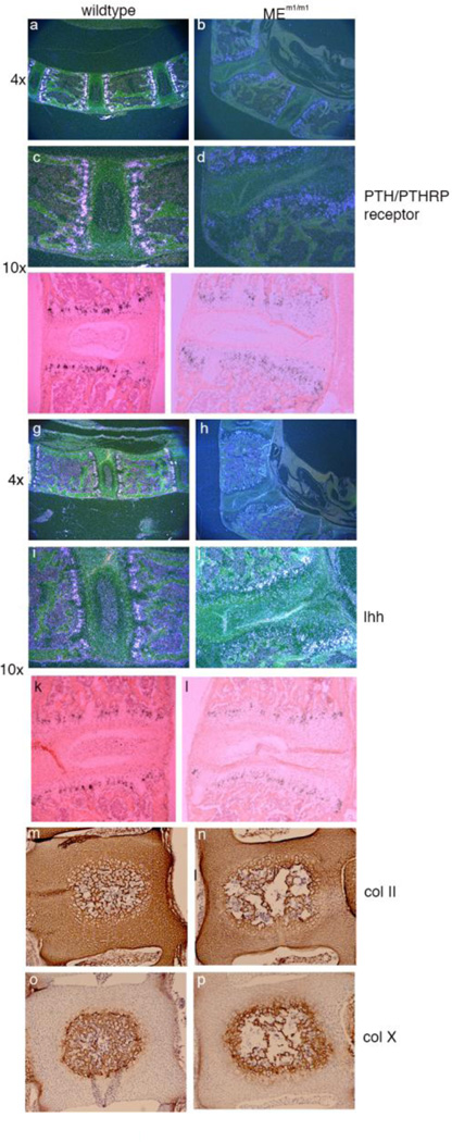
Figure 6. Analysis of endochondral bone formation. Panels a-l.
In situ hybridization of adult lumbar spine for Prp (PTH/PTHRP receptor) (panels a-f; a-d, dark field; e, f, bright field) and Ihh (Panels g-l; g-j, dark field; k, l, bright field). Original magnification is given to the left. Panels m-p. Immunohistochemistry for collagen 2a1 and collagen 10a1 in newborn wildtype and MEm1/m1 mice. Coronal sections of L4 vertebrae in paraffin. Panels m, n: collagen 2a1 stain; Panels o and p: collagen 10a1 stain. Collagen 2 is expressed throughout the early vertebral body and IVD, while collagen 10a1 is limited to the mineralizing core of the vertebral body.

