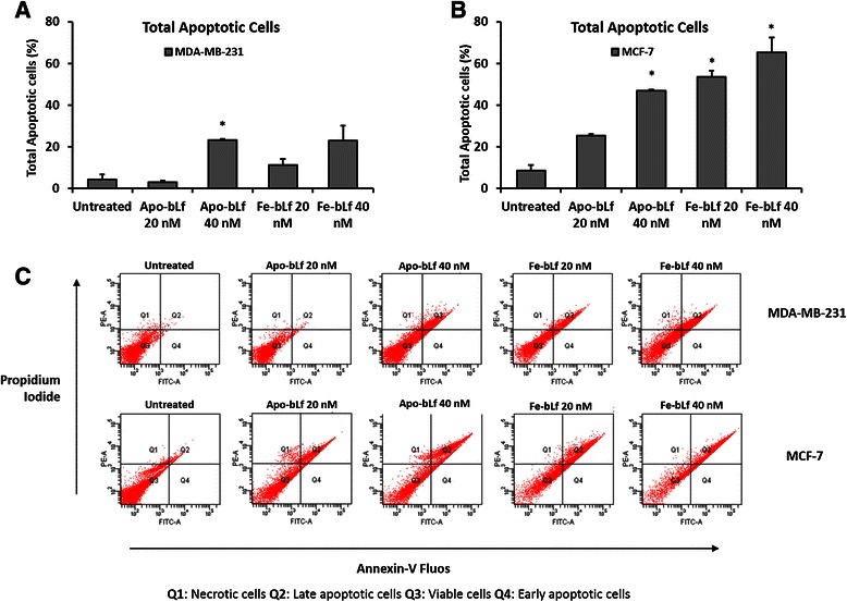
Fig. 4.

BLf-induced apoptosis in MDA-MB-231 and MCF-7 cells. Annexin-V-Fluos staining detected apoptosis in MDA-MB-231 and MCF-7 cells following bLf treatments. Cells were treated for 24 h with Apo-bLf and Fe-bLf at concentrations of 20 nM and 40 nM. a Total apoptotic MDA-MB-231 and b MCF-7 cells following bLf treatments. Flow cytometry plots of MDA-MB-231 and MCF-7 cells (c). Q1: Necrotic cells, Q2: Late apoptotic/Dead cells, Q3: Viable cells, Q4: Early apoptotic cells. Total apoptotic cells were the sum of early and late apoptotic cell populations. Data represented as means +SEM, student t-test used for statistical analysis
