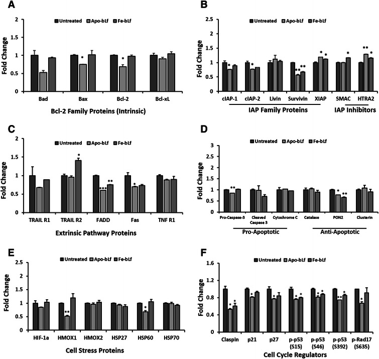
Fig. 5.

Apoptosis protein array MDA-MB-231 breast cancer cells. Apoptosis protein array results following incubation of MDA-MB-231 cell lysate after treatments with Apo-bLf and Fe-bLf at 40 nM for 24 h. Cell lysate (250 μg) incubated with nitrocellulose membrane pre-labelled with capture antibodies (duplicate spots) and detected via chemiluminescence. a Bcl-2 family proteins Bad, Bax, Bcl-2 and Bcl-xL. b Inhibitor of apoptosis (IAP) proteins cIAP-1 and 2, Livin, Survivin and XIAP, and inhibitors SMAC and HTRA2. c Extrinsic pathway proteins and receptors TRAIL 1 and 2, FADD, Fas and TNF R1. d Pro-apoptotic proteins Pro-caspase-3, Cleaved Caspase-3 and Cytochrome C, anti-apoptotic proteins Catalase, PON2 and Clusterin. e Cell stress proteins HIF-1α, HMOX1, HMOX2, HSP27, HSP60 and HSP70. f Claspin, p21, p27, phospho-p53 (S15), phospho-p53 (S46), phospho-p53 (S392) and phospho-Rap17 (S635). Average density determined using ImageJ software and fold change calculated compared with untreated. Data represented as means with + SEM. * = p < 0.05, ** = p < 0.01, *** = p < 0.001 compared with the untreated group. Statistical analysis was performed via Student’s t-test
