
Figure 8.
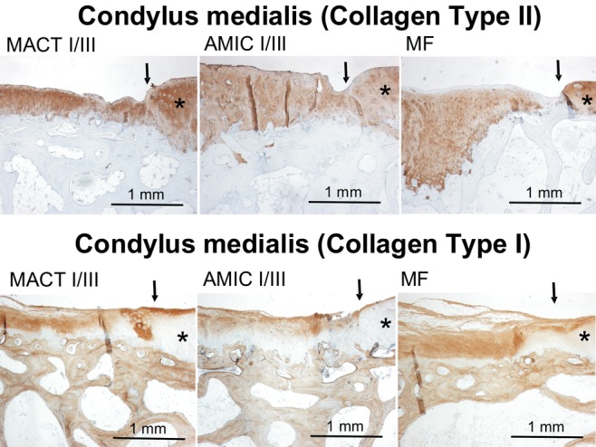
Examples of immunohistochemical collagen type II and type I staining in condylar defects of different experimental groups. Collagen expression is indicated by brown staining. There is no collagen type II staining in subchondral bone, while bone tissue is positively stained for collagen type I, giving an internal positive control. Arrows mark the borders of the defects; stars indicate more or less intact cartilage tissue surrounding the defect area. Because of the high variance in staining, these examples do not represent a mean intensity of staining of the corresponding experimental group but give an impression of the quality of staining in general.
Note: MACT I/III = matrix-associated autologous chondrocyte transplantation + Chondro-Gide scaffold; MF = microfracture; AMIC I/III = autologous membrane-induced chondrogenesis + Chondro-Gide scaffold.