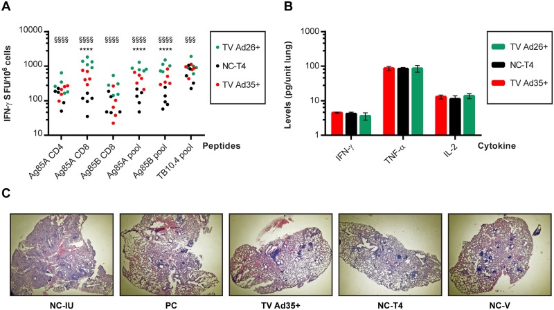
Fig 7. Probing the mechanism of failure at week 28.
A) ELISpot analysis was conducted at the final relapse time point of week 28 as previously described. Results of groups that received immunotherapy (TV Ad35+ and TV Ad26+) and the main negative control group NC-T4 are depicted. Results of significance testing are depicted by * for comparison between TV Ad35+ and NC-T4 whereby: * p<0.05, ** p<0.01, *** p<0.001 and **** p<0.0001. § denotes significant differences between TV Ad26+ and NC-T4 whereby: § p<0.05, §§ p<0.01, §§§ p<0.001 and §§§§ p<0.0001. Statistical significance was determined by ANOVA followed by Tukey’s multiple comparisons test where n = 5. B) Multiplex enzyme-linked immunosorbent assay was carried out at week 28 on lung samples as previously described. Results from groups that received immunotherapy (TV Ad35+ and TV Ad26+) and the main control group NC-T4 are shown (mean and standard deviation, n = 4 per group). No statistically significant differences in the levels of IFN-γ, TNF-α or IL-2 were measured between all groups (IFN-γ: p = 0.35; TNF-α: p = 0.98; IL-2: p = 0.09; ANOVA). C) Histopathology of lung samples at week 12 was examined for signs of immunopathology following two doses of immunotherapy with Ad35-TBS. In comparison to the NC-IU animals, the lungs of animals that underwent therapy appeared similar with minimal signs of lesions, necrosis or significant immune infiltration. Immunotherapy did not result in observable immunopathology. Results of histopathology at week 12 are representative of all time points measured (weeks 4, 6, 8 and 10) and at week 18 following Ad26-TBS boosting (data not shown).

