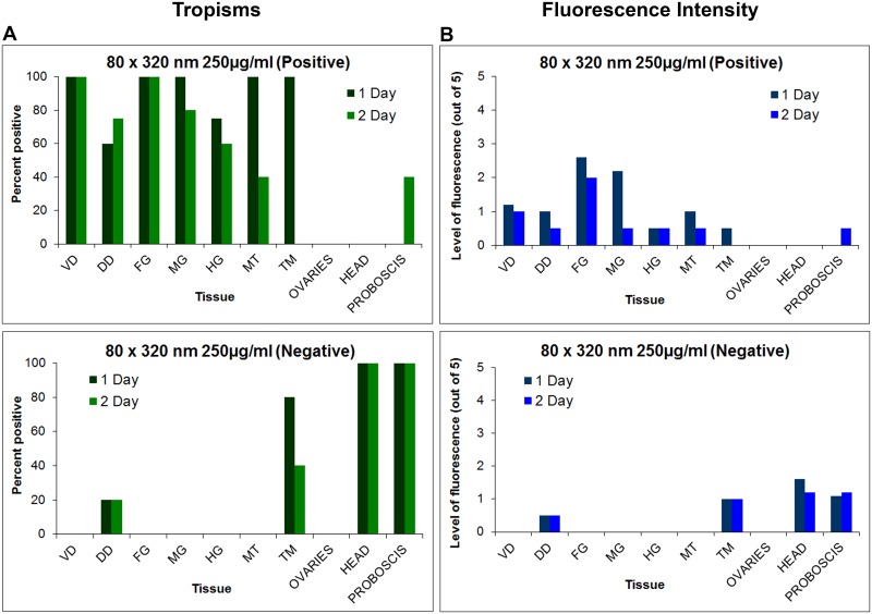
Fig 11. Tissue tropisms and fluorescence intensity of positively and negatively charged 80 nm x 320 nm hydrogel nanoparticles in Anopheles gambiae females following parenteral challenge.
(A) Tissue tropisms (percent of mosquitoes with NP fluorescent signal of any intensity detected in the respective organ or tissue) of positively and negatively charged 80 x 320nm NPs in An. gambiae following parenteral challenge (250 μg/ml). (B) Fluorescence intensity (mean level of fluorescence intensity (NP load) in organs and tissues containing NPs) of positively and negatively charged 80 x 320 nm NPs in An. gambiae organs and tissues following parenteral challenge. NP tissue tropisms and loads were greater and of longer duration in most tissues females injected with positively charged NPs than in those injected with negatively charged NPs. Negatively charged NPs were most frequently associated with head and proboscis tissues. VD = ventral diverticulum; DD = dorsal diverticula; FG = foregut; MG = midgut; HG = hindgut; MT = Malpighian tubules; TM = thoracic muscle.

