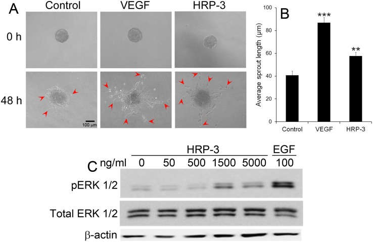
Fig 5. HRP-3 induces endothelial cell sprouting and ERK activation.
(A) Endothelial cell sprouts induced by HRP-3. HUVEC spheroids were embedded in fibrin gel and cultured in EBM-2 medium in the presence of HRP-3 (10 ng/ml), VEGF (2.5 ng/ml) or PBS for 48 h. Bar = 100 μm. (B) Quantification of endothelial cell sprouts. The average length of sprouts per spheroid was quantified as described in S1 Fig. A total of 10 spheroids per group were quantified (n = 10). Data are mean ± s.e.m. in one representative experiment. **P<0.01, *** P<0.001, vs. control. (C) HRP-3 activates ERK pathway. HUVECs were starved in serum-free medium for 45 min, followed by incubation with HRP-3 or EGF (positive control) for 10 min. The cell lysate were analyzed by Western blot using antibody against ERK, phospho-ERK (pERK) or β-actin. Both experiments were validated three times with similar outcomes.

