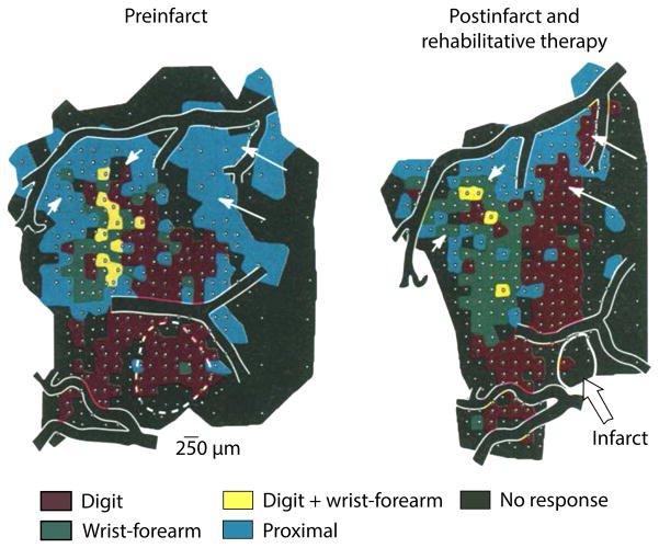
Fig. 1. Why Ipsilesional Primary Motor Cortex (M1) was first targeted with stimulation in rehabilitation.
Adapted from Nudo and others (1996). Ipsilesional M1 was first targeted with stimulation in stroke rehabilitation because pioneering work in animal models had suggested that the region shows adaptive plasticity with return of skill in recovery. Here, Nudo and others (1996) illustrate how ipsilesional M1 reorganizes with rehabilitative training in a non-human primate model of stroke. The infarct (dashed circle) has destroyed ~22% of the digit (red) representations and 4% of the wrist-forearm (green) representations in the region of M1. With rehabilitative training involving the distal forelimb, the animal exhibits reorganization of representations within peri-infarct M1 (right). The spared digit representation expands by ~15% while the wrist-forearm representations increase by 58.5%. Nudo and others conclude that with skill relearning, surviving representations of trained distal forelimb expand in the peri-infarct territory, as well as occupy territory previously claimed by proximal segments (indicated by white arrows denoting areas in blue). These pivotal findings emphasized two important themes in stroke recovery: skill-based rehabilitative training invokes adaptive plasticity in the stroke brain and peri-infarct M1 lies at the center of this process. Thus, subsequent studies chose peri-infract M1 as a target for stimulation in rehabilitation.

