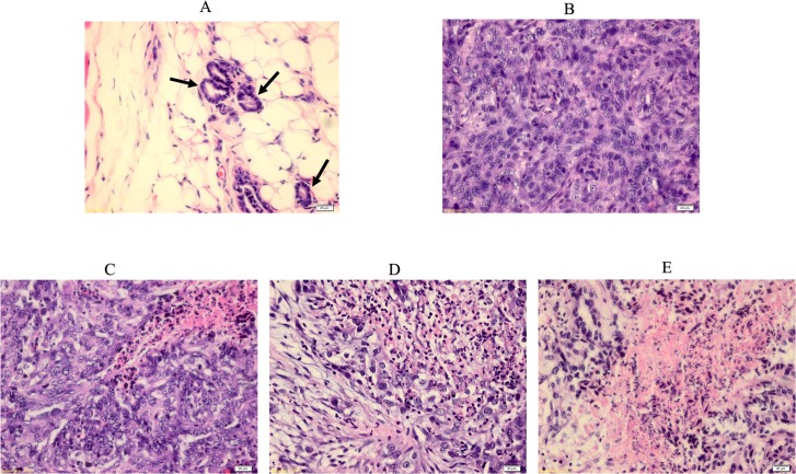
Fig 3. H & E staining of normal and breast cancer tissues.
Histopathological observation of normal and tumor sections represents the mammary tissues from various experimental rat groups. Arrow shows a breast normal duct. (A), LA7-induced breast tumor (B), FLAHE low-dose treatment (C), FLAHE high-dose treatment (D) and tamoxifen-treated group (E). Histological examination of normal breast and tumor breast cancer before and after FALHE treatment. The normal breast shows normal duct tissues, but the LA7-induced breast tumor shows a disruption in morphology and an invasion of ductal cells throughout the breast tissues. Magnification, 40×.

