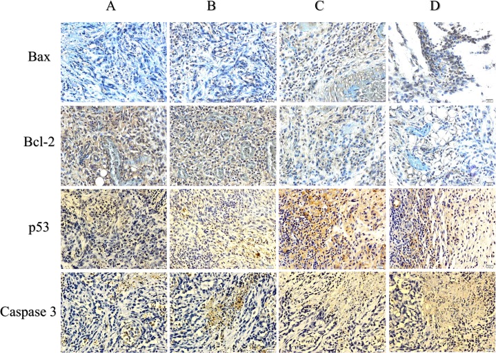
Fig 6. Immunohistochemical results for Bcl-2, Bax, p53 and caspase 3.
Tumor control (A), FALHE low-dose treatment (B), FALHE high-dose treatment (C), and tamoxifen treatment (D). Dark brown particles are representing the expression of Bax, Bcl-2, Caspase 3 and p53 proteins. Microscopic observation of the FALHE-treated group compared with the control tumor showing high expression levels of Bax, p53 and caspase 3 and a low expression level of Bcl-2 protein. This result demonstrates the activation of the intrinsic pathway of apoptosis. Magnification, 40×.

