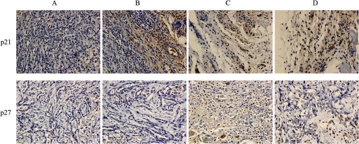
Fig 7. Immunohistochemical results for p21 and p27.
Various groups of mammary tumor were screen as: Tumor control (A), FALHE low-dose treatment (B), FALHE high-dose treatment (C), and tamoxifen treatment (D). Dark brown particles are indicating the expression of p21 and p27. High expression of p21 and p27 was observed after FALHE treatment compared with the tumor control, revealing cell cycle arrest.

