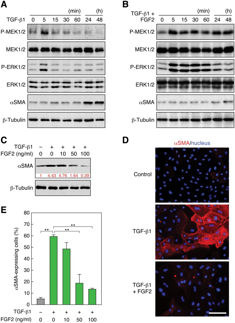
Fig 1. FGF2 induces sustained activation of the Ras—ERK pathway and inhibits TGF-β1-induced EMT.
(A) Transient phosphorylation of MEK and ERK and induction of αSMA expression by TGF-β1 stimulation. RLE cells were stimulated with 0.5 ng/ml TGF-β1. The levels of MEK, phospho (P)-MEK, ERK, P-ERK, and αSMA, as well as β -tubulin as a standard, were analyzed by immunoblotting. (B) Sustained phosphorylation of MEK and ERK and inhibition of αSMA expression by FGF2 stimulation. RLE cells were stimulated with 100 ng/ml FGF2 in combination with 0.5 ng/ml TGF-β1. (C) A dose-dependent reduction of the TGF- β 1-induced αSMA protein level by FGF2 stimulation. RLE cells were stimulated with the indicated concentrations of FGF2 together with 0.5 ng/ml TGF- β 1 for 48 h. The relative intensity of αSMA band is indicated under the blot. (D) Induction of αSMA expression by TGF- β 1 and suppression of the expression by FGF2. RLE cells were stimulated with 0.5 ng/ml TGF- β 1 or with 100 ng/ml FGF2 along with TGF- β 1 for 48 h. αSMA expression and localization was detected by immunofluorescent staining with the Cy3—anti-αSMA mAb (red) as well as nuclear staining with Hoechst 33258 (blue). Scale bar, 50 μm. (E) A dose-dependent reduction of the ratio of TGF- β 1-induced αSMA-expressing cells by FGF2 stimulation. αSMA-expressing cells were detected as in (D). The values are means ± SD of 3 independent experiments. **, P < 0.01 by t test.

