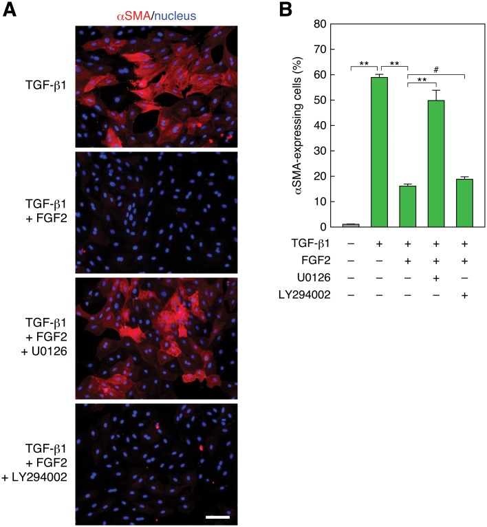
Fig 2. Inhibition of MEK but not PI3K recovers TGF-β1-induced and FGF2-suppressed EMT.
(A) Recovery of FGF2-suppressed αSMA expression by MEK inhibition but not by PI3K inhibition. RLE cells were pretreated with 10 μM of the MEK inhibitor U0126 or the PI3K inhibitor LY294002 for 30 min. Then they were stimulated with 0.5 ng/ml TGF-β1 along with 100 ng/ml FGF2 for 48 h. αSMA expression (red) and nuclei (blue) were detected as described in Fig 1 legend. Scale bar, 50 μm. (B) The ratio of αSMA-expressing cells in the analysis of (A). The values are means ± SD of 3 independent experiments. **, P < 0.01; #, P > 0.05 (not significant) by t test.

