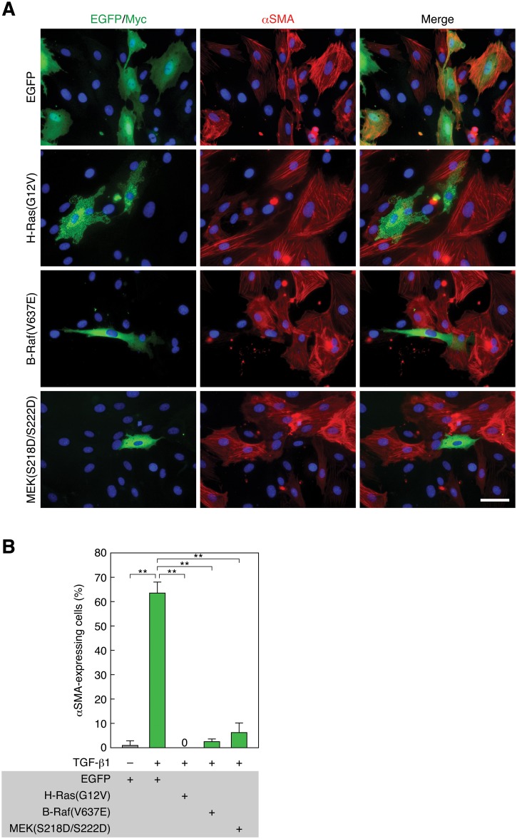
Fig 3. Constitutively active H-Ras, B-Raf, or MEK1 suppresses TGF-β1-induced EMT.
(A) Suppression of TGF-β1-induced EMT by constitutively active H-Ras, B-Raf, and MEK1. RLE cells were transfected with Myc-tagged H-Ras(G12V), EGFP-tagged B-Raf(V637E), EGFP—MEK1(S218D/S222D), or EGFP expression vector. Twenty-four hours after the transfection, they were treated with 0.5 ng/ml TGF-β1 for 48 h. αSMA expression (red) and nuclei (blue) were detected as described in Fig 1 legend. Myc- and EGFP-tagged proteins were detected by anti-Myc pAb and anti-GFP pAb staining, respectively (green). Scale bar, 20 μm. (B) The ratio of αSMA-expressing cells in the analysis of (A). The values are means ± SD of 3 independent experiments. **, P < 0.01 by t test.

