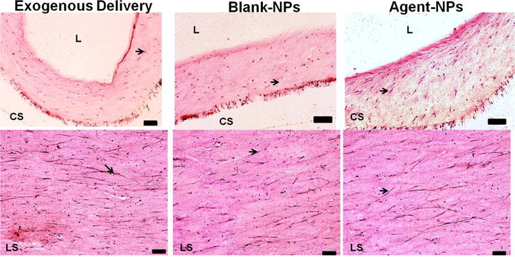
Figure 7.

Elastic staining of 30 μm thick cross- (CS; upper panel) and longitudinal sections (LS; lower panel) of collagen constructs (stained pink) after 21 days of treatment. Orientation of cells and elastic fibers (stained purple/black, indicated by arrows) were seen in the longitudinal direction. (Scale bars for upper panel are equivalent to 100 μm; scale bars for lower panel equivalent to 500 μm)
