
Figure 1. Model of the pathogenesis of autoimmune valvular carditis.
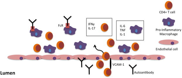
CD4+ T cells, with the help of VCAM-1, and macrophages extravasate across the endothelium into the valve interstitium. Th1 and Th17 effector CD4+ T cell lineages are characterized by the production of IFNγ and IL-17, respectively. Macrophages, in some cases stimulated by antibodies engaging activating Fc receptors, release the pro-inflammatory cytokines IL-6, TNF, and IL-1.