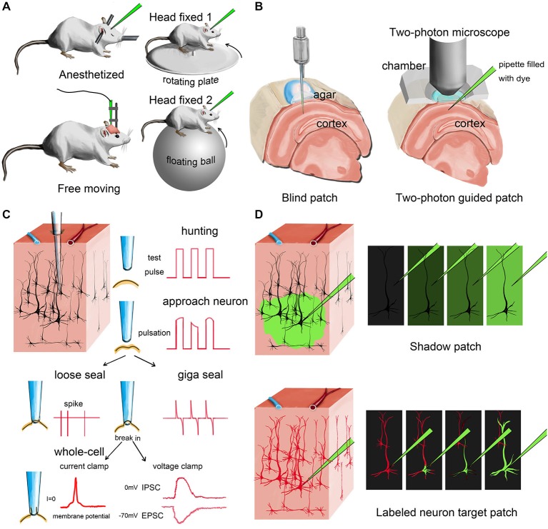
Figure 1.
Different modes of in vivo patch-clamp recording. (A) Representative in vivo patch-clamp setups for anesthetized, awaking and behaving animals. (B) Demonstration of blind patch and two-photon-guided patch. (C) Procedures and different recording modes of in vivo patch clamp (blind patch). When the pipette approaches a nearby cell, heartbeat-associated changes become notable in test pulses. Releasing positive pressure allows the pipette tip to form a loose seal or a giga seal for loose-patch recording or whole-cell recording, respectively. After giga seal formation, the cell membrane can then be broken for either current-clamp recording or voltage-clamp recording. (D) Two different methods for visually guided in vivo patch clamp: shadow patch and labeled-neuron-guided patch.

