Fig. 2.
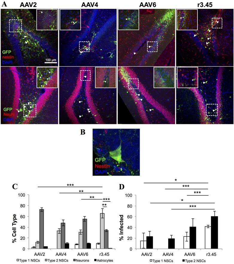
Selectivity towards and infectivity of adult NSCs in the rat brain. (A) Representative images at low (main, 20×) and high (inset, 100×) magnification of the rat dentate gyrus 3 weeks post-injection of recombinant AAV2, AAV4, AAV6 or AAV r3.45 vectors expressing GFP (green). Brain sections were co-stained for nestin (top row, red) or NeuN (bottom row, red) along with DAPI (blue), and infected cells of each type are marked with arrowheads. Dashed rectangles indicate the regions magnified in the insets. Scale bar: 100 μm. (B) Representative image of a Type 1 NSC infected with AAV r3.45 vector. (C) The percentage of GFP+ cells co-staining for markers of each cell type was quantified to determine the selectivity of each viral vector. (D) The percentage of nestin+/Gfap+ (Type 1) or nestin+/Sox2+ (Type 2a) cells infected by each viral vector was quantified to determine NSC infectivity. Error bars indicate s.d. (n=3); *P<0.05, **P<0.01, ***P<0.005 (ANOVA).

