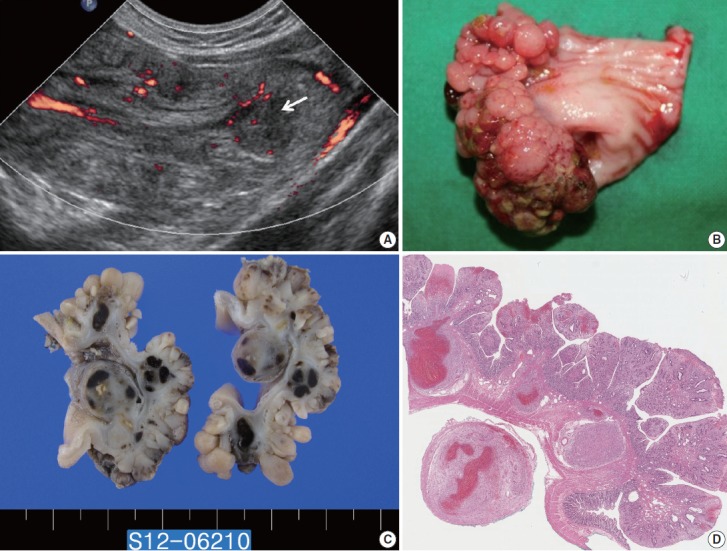
Fig. 1.

(A) Longitudinal ultrasonography of the lower abdomen with a Doppler study shows an ovoid mass with alternating thick hypoechoic and thin hyperechoic layers, indicating ileoileal intussusception and Doppler flow signals at the intussusceptum. A round hypoechoic lesion (arrow) indicating a lead point of intussusception is identified. (B) The ileum reveals a 4×2×1 cm, roughly ovoid, sessile, polypoid mass with a conglomerated nodular or nodule-aggregating appearance. (C) The cut surface shows thickened mucosa and multiple round solid nodules with focal hemorrhages at deeper layers. (D) The polyp is composed of enlarged plicae circulares having dilated and distorted crypt glands with expanded lamina propria.
