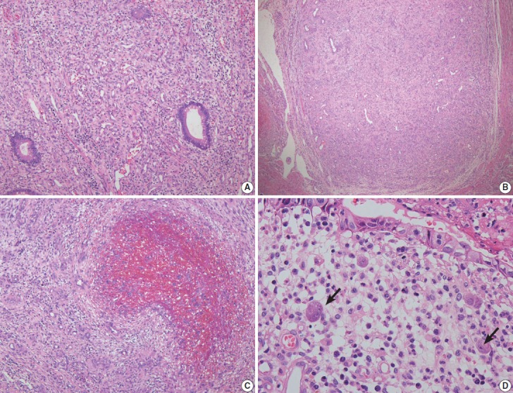
Fig. 2.

(A) The lamina propria shows granulation tissue-type small vessel proliferation. (B) The nodules at deeper layers are composed of proliferated florid small vessels and fibroblasts. (C) Organizing-thrombus-like areas are noted focally in the nodules. (D) A few stromal cells with intranuclear and cytoplasmic cytomegalovirus inclusions are present beneath the eroded mucosal surface.
