
Figure 4. Sox2+ Cells Propagate Ptc MBs In Situ.
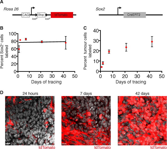
(A) To perform lineage tracing in MB, mice with a loxP-stop-loxP tdTomato reporter gene at the Rosa 26 locus and Sox2creER knock in mice were crossed to the Ptc model.
(B) Quantification of the frequency of cells labeled with tdTomato within the Sox2+ tumor population following a 5 mg tamoxifen injection (n = 3–5 per time point, mean ± SEM).
(C) Quantification of tdTomato labeling of tumor cells following a 5 mg tamoxifen injection (n = 4–6 per time point, mean ± SEM).
(D) Representative images of tumor labeling with tdTomato at 24 hr, 7 days, and 42 days post-tamoxifen. DAPI is shown in white. Scale bar, 11 μm.
See also Figure S4.