Figure 4.
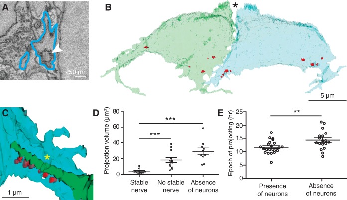
Retraction of projections is associated with stable afferent contact. (A) An SBEM image shows an immature synaptic ribbon (arrowhead) within a hair cell projection (blue outline). (B) In a reconstruction of the sibling hair cells from a recent division, synaptic ribbons (red) cluster at the sites from which projections emerge. N = 10. In this basal view, the partially developed hair bundles are apparent at the cells’ apical ends (asterisk). (C) An afferent terminal of the functionally appropriate subpopulation (green) contacts immature synaptic ribbons (red) on a hair cell projection (blue). A sheet of hair cell membrane (asterisk) folds over the afferent terminal. (D) Enlarged projections are associated with the absence of stable nerve contacts (P < 0.001) or of lateral line neurons (P < 0.0001). N = 10, 12, and 11. (E) The total period during which projections emerge from hair cells increases in the absence of lateral line neurons. P < 0.01; N = 23 and 18.

