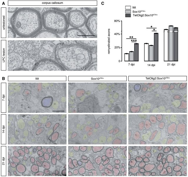
Figure 7.
Gain of Olig2 function in adult OPCs accelerates remyelination. (A) Electronic micrographs of unlesioned and LPC demyelinated corpus callosum. In unlesioned corpus callosum, axons of small calibres are unmyelinated. In demyelinated lesions, remyelinated axons are characterized by a thin myelin sheath. (B) Quantification of remyelinated axons in lesions of doxycycline treated wild-type (white bars), Sox10rtTA/+ (light grey bars) and TetOlig2:Sox10rtTA/+ (dark grey bars) mice at 7, 14 and 21 days post-injection (dpi). The percentage of remyelinated axons was significantly increased in TetOlig2:Sox10rtTA/+ at 7 and 14 days post-injection, compared to wild-type (P7dpi = 0.0095, P14dpi = 0.0413) or Sox10rtTA/+ mice (P7dpi < 0.001, P14dpi = 0.0343). However at 21 days post-injection, the percentage of remyelinated axons was similar in the three groups. (C) Electron micrographs illustrating remyelination of the lesions in wild-type, Sox10rtTA/+ and TetOlig2:Sox10rtTA/+ mice at 7, 14 and 21 days post-injection. Remyelinated, demyelinated and non-demyelinated axons were colour-coded in red, yellow and blue, respectively. Scale bars: A = 500 nm; C = 2 μm.

