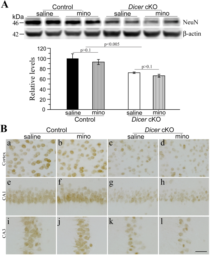
Figure 3. Minocycline did not ameliorate neuron loss in Dicer cKO mice.
(A) Western blotting for NeuN using cortical lysates. β–actin served as the internal control. In the bar graph, there was significant difference in NeuN levels between control and cKOs. NeuN levels were not different between saline- and minocycline- treated Dicer cKOs. (B) Immunohistochemistry for NeuN. Dicer cKO mice (c,d,g,h,k,l) exhibited less number of NeuN+cells than control animals (a,b,e,f,i,j) did. However, there was no difference in the number of NeuN+cells between minocyline- and saline-treated Dicer cKOs. Scale bar = 40μm. Raw Western blotting images for NeuN were shown in Supplementary Figure 4.

