
Figure 1.
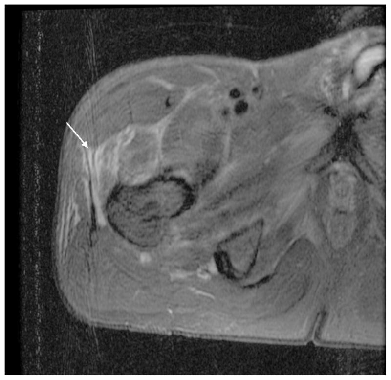
MRI of hip revealing increased T2 signal intensity along the fascial planes in the vicinity of right hip, involving anterior upper thigh, the adductor group, gluteus minimus, and the tensor fascia lata (white arrow) suggesting myositis with no evidence of abscess, soft tissue gas, or septic hip or osteomyelitis.