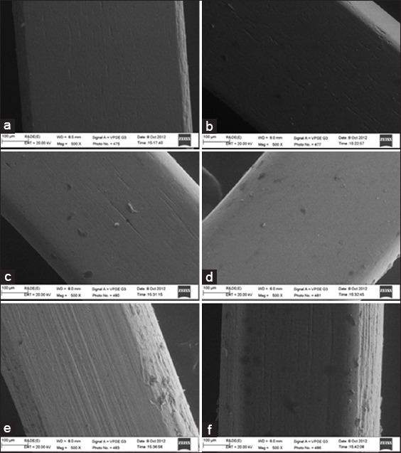
Figure 4.

(a) Scanning electron microscope (SEM) image for commercially available uncoated stainless steel (SS) wire (0.019” × 0.025”). (b) SEM image for nanoparticle coated SS wire (0.019” × 0.025”). (c) SEM image for commercially available uncoated nickel-titanium (NiTi) wire (0.019” × 0.025”). (d) SEM image for nanoparticle coated NiTi wire (0.019” × 0.025”). (e) SEM image for commercially available uncoated titanium molybdenum alloy (TMA) wire (0.019” × 0.025”). (f) SEM image for nanoparticle coated TMA wire (0.019”× 0.025”).
