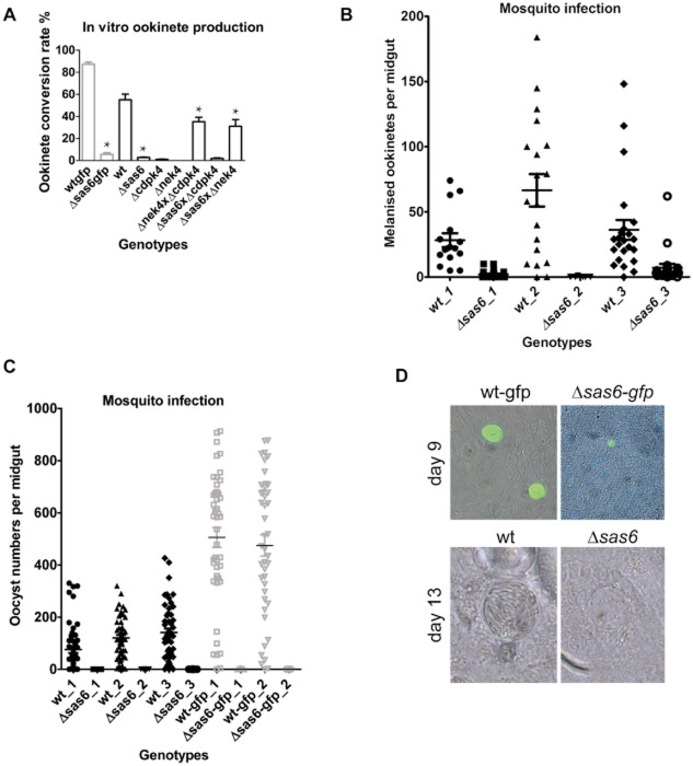
Fig 5.
Analysis of sexual reproduction and infectivity to mosquitoes in Δsas6 and wt clones.A. Ookinete conversion rate as measured by the number of ookinetes per total number of fertilized and unfertilized females in Δsas6-gfp, Δsas6 and crosses of Δsas6 with Δcdpk4 (male deficient) and Δnek4 (female deficient) clones. Δsas6 form significantly fewer ookinetes than wt, a phenotype that can be rescued by crossing it with Δnek4. Asterisk * indicates statistically significant differences in Student's t-test with P-values lower than 0.01, Table S1.B. Ookinete invasion of An. gambiae L3-5 midguts. Mosquitoes were infected with wt and Δsas6, mosquitoes were dissected at day 6 and melanized ookinetes were counted in midguts in 3 different biological replicates. Intensity of infection is significantly decreased in all replicates, Table S2A.C. Infectivity of Δsas6 to An. stephensi mosquitoes. Mosquitoes were infected with wt, wt-gfp, Δsas6 and Δsas6-gfp, midguts were dissected at day 12 and oocysts were counted in 3 different biological replicates. Intensity and prevalence of infection are significantly different from wt, Table S2B.D. Bright-field and fluorescent oocyst images at day 9 and day 13. At both time points, Δsas6 appear smaller than wt. This size difference was quantified and is statistically significant at day 9 (Table S1). At day 13, wt oocysts display densely packed slender sporozoites that appear like striations, in contrast Δsas6 oocysts appear empty.

