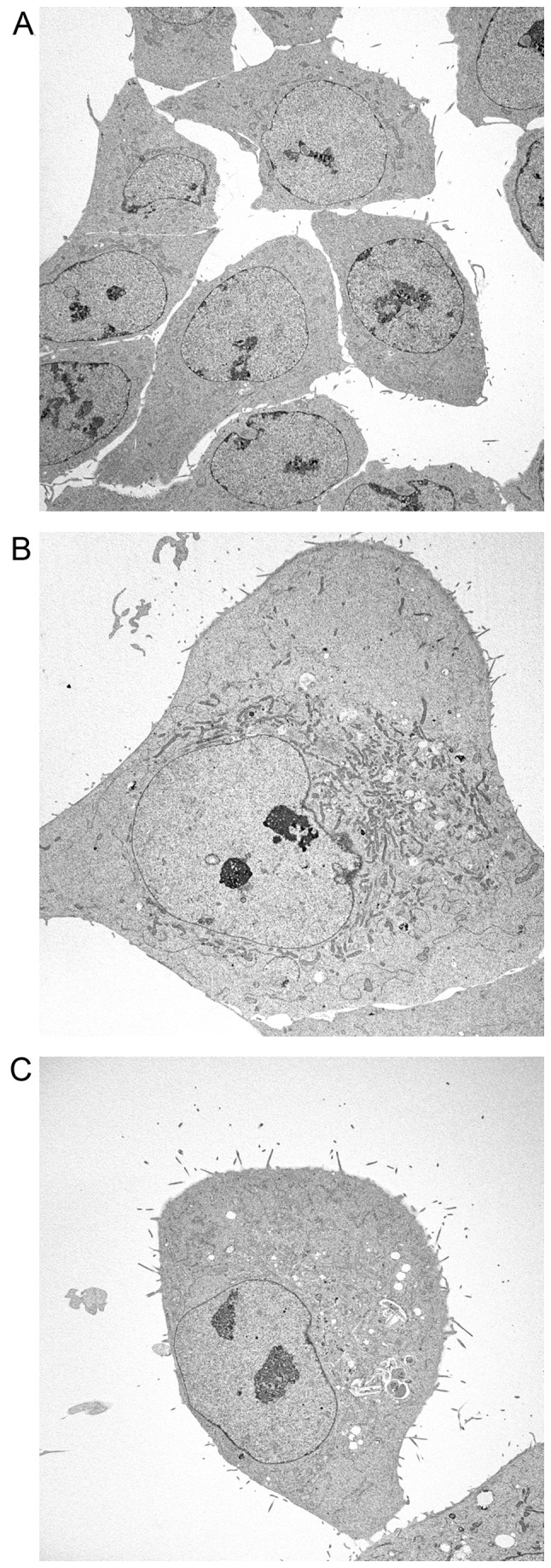
Figure 5.

Electron microscopy analysis of untreated HCT116 cells (A), or HCT116 cells treated with FTD (B) or FdUrd (C). Cells were treated in 60-mm culture dishes with 6 μmol/l FTD or 3 μmol/l FdUrd for 72 h, using a concentration found capable of 50% growth inhibition (data not shown) and subsequently fixed. After dehydration by ethanol treatment, samples were transferred to resin and polymerized. The acceleration voltage of an electron microscope was set at 80 kV, and observations were performed under ×714 magnification.
