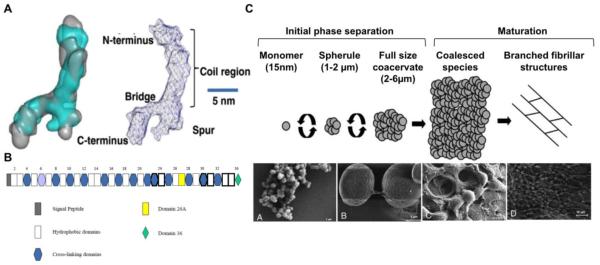
Figure 2. Tropoelastin structure and assembly.
(A) Tropoelastin molecule structure obtained by superimposing filtered average small angle x-ray scattering (SAXS) and small angle neutron scattering (SANS) models (left) and labeled diagram of the model for full-length tropoelastin showing proposed locations of the N-terminus, the spur region containing exons 20–24, and the C-terminus (Scale bar, 5 nm). Adapted from[40]. (B) Human tropoelastin domain map, showing all exons. Exons subject to alternative splicing are outlined in bold. Exon 26A is rarely expressed in human tropoelastin. Domain 36 is uniquely designated because of its distinct structural features. Adapted from [42]. (C) Stages of in vitro tropoelastin self-assembly. The initial phase separation is a reversible process characterized by the formation of 1–2 μm spherules which progressively grow into ~6 μm assemblies. The second maturation stage involves irreversible aggregation of full-sized coacervates into larger species which may display branched fibrillar structures. Scanning electron micrograph of tropoelastin spherules (A), full-sized coacervates (B), and coalesced species (C–D). Adapted from [46, 47].

