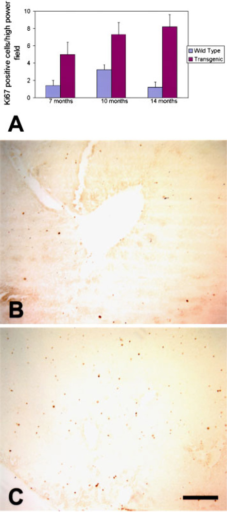
Figure 3.
Increased proliferation in transgenic livers. The proportion of proliferating cells is increased in livers from CMV/Cux-1 mice (A). The data are plotted as the number of Ki67 positive cells per high power field. Ki67 positive cells were counted in five high power fields from four different animals at each time point. Cell counts in transgenic animals were obtained from extrafocal regions. Sections are shown from 10-month-old wild type liver (B) and extra-focal region of transgenic liver (C). Standard deviation is indicated by error bars. Original magnification: 100× (Bar, 5 µm). [Color figure can be viewed in the online issue, which is available at www.interscience.wiley.com.]

