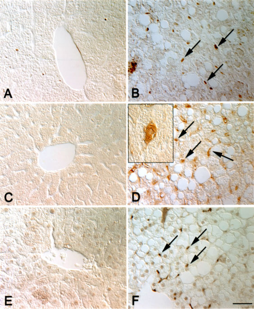
Figure 4.
Increased proliferation of hepatocyte precursor cells in transgenic livers. Wild type (A, C, E) and transgenic (B, D, F) liver sections from 8-month-old mice were labeled with antibodies directed against Cux-1 (A, B), chromogranin-A (C, D), or PCNA (E, F). Cux-1 was ectopically expressed in small cells (arrows in B) between the hepatocytes in transgenic livers, but was not detected in wild type livers (A). Transgenic livers showed an increase in the number of cells labeled positively for chromogranin-A (arrows in D), a marker for hepatocyte precursor cells. Inset in (D) shows that chromogranin-A was detected in cells morphologically resembling hepatocytes, indicating differentiation to the hepatocyte lineage. No chromogranin-A positive cells were observed in the wild type liver (C). The small, fibroblastic shaped cells, but not the hepatocytes, labeled positively for PCNA (arrows in F), indicating that they are proliferating. No PCNA positive cells were detected in the wild type liver (E). Original magnification: 400× (Bar, 50 µm). [Color figure can be viewed in the online issue, which is available at www.interscience.wiley.com.]

