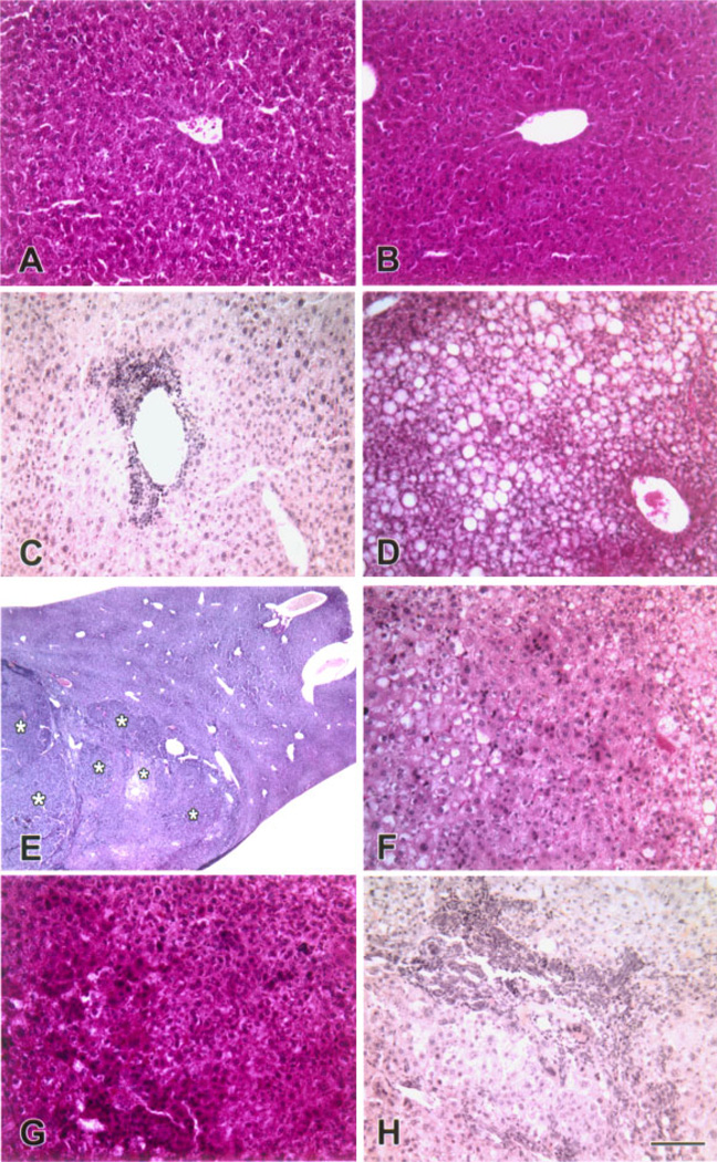
Figure 7.
Progression of liver lesions in CMV/Cux-1 transgenic mice. Light micrographs of wild type (A) and transgenic livers (B-H). At 6 months of age, transgenic livers (B) are indistinguishable from wild type livers (A). At 8 months of age, non-suppurative inflammation (C) and fatty cell change (D) are observed in transgenic livers. At 10 months of age, multifocal lesions are evident in transgenic livers (asterisks in E). Higher magnification reveals that these are of the mixed cell type (F). At 14 months of age, hepatocellular carcinoma is present in transgenic livers (G). Biliary hyperplasia is also observed (H). Original magnification: (A–D, F–H) 200× (Bar, 100 µm). (E) 13×. [Color figure can be viewed in the online issue, which is available at www.interscience.wiley.com.]

