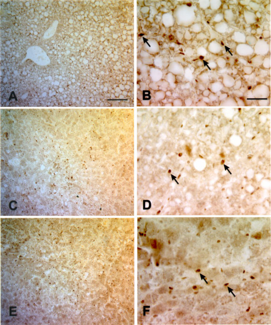
Figure 9.
Ectopic expression of Cux-1 increases in lesion progression. Localization of Cux-1 protein in 8-, 10-, and 14-month-old transgenic livers showing fatty cell change (A, B), mixed cell foci (C, D), and hepatocellular carcinoma (E, F). Small Cux-1 positive cells were observed throughout the 8-month-old transgenic livers in areas of fatty cell change (A, and arrows in B). In some areas of 10-month-old transgenic liver, corresponding to mixed cell foci, high concentrations of Cux-1 positive cells were observed (C). Higher magnification revealed that the Cux-1 positive cells corresponded to only small cells (arrows in D), while hepatocytes were not labeled for Cux-1. In a section of 14-month-old transgenic liver corresponding to hepatocellular carcinoma, Cux-1 positive cells were observed (E), and higher magnification revealed that these cells were small cells (arrows in F), but not hepatocytes. Original magnification: (A, C, E) 100× (Bar, 100 µm). (B, D, F) 400× (Bar, 50 µm). [Color figure can be viewed in the online issue, which is available at www.interscience.wiley.com.]

