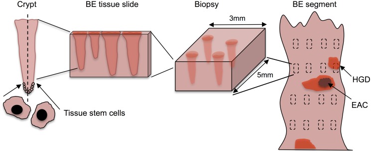
Fig 1. The multiscale nature of BE screening.
Biopsy screening for BE scales from stem cells in the crypt (left) to the BE cylindrical segment of the esophagus depicted (right) with rectangles representing biopsy samples taken during endoscopy via the Seattle biopsy protocol. The BE segment may have dysplasia and/or malignant tissue patches that remain untouched after biopsying. During histological preparation, portions of each biopsy are sliced by microtome and placed on slides for pathologic assessment. Diagnosis is made by microscopic interpretation of crypt and cellular architecture, reflecting the most severe tissue grade found from all slides.

