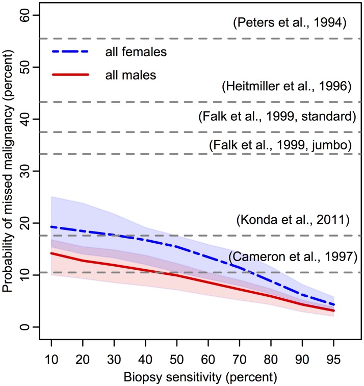
Fig 5. Predicted probability of missed malignancy in positive high grade dysplasia population at index screen.
Percentages of patients diagnosed with HGD during index endoscopy (denominator is population plotted in Fig 4) who concurrently harbored missed, malignant clone(s) present on their BE segments that were not detected on biopsy screen. Since the sensitivity of each study is unknown, literature values for the corresponding probability of missed malignancy are depicted as horizontal grey dotted lines at a single percentage level [25, 30–34]. One publication included percentage of occult malignancy after HGD was diagnosed with either standard or jumbo forcep sizes, as indicated [38]. Expected proportions produced with 100K BE patient simulations each for males (red, solid) and females (blue, dash-dotted) with assumed density of σ = 3300 stem cells/mm2 (shaded regions represent sensitivity of results for σ ∈ [2000, 5000]).

