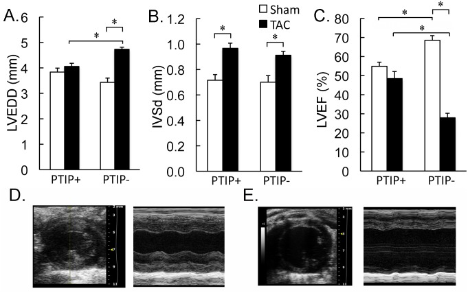
Fig 3. LV chamber dilation and depressed cardiac function in PTIP- TAC hearts.
Echo performed on PTIP+ hearts after sham (n = 9) and TAC (n = 10) revealed a significant increase in anterior wall thickness (IVSd; panel B) after TAC without any significant change in LV end diastolic diameter (LVEDD; panel A) or LV ejection fraction (LVEF; panel C). PTIP- hearts after TAC (n = 12) also demonstrated an increase in anterior wall thickness compared with PTIP- sham mice (n = 10). However, PTIP- TAC hearts reveal a significant increase in LVEDD (panel A) and a significant decrease in LVEF when compared to PTIP- sham and PTIP+ TAC hearts. Representative 2D and M-mode images are shown for PTIP+ TAC (panel D) and PTIP- TAC (panel E). Data shown are means ± SEM.

