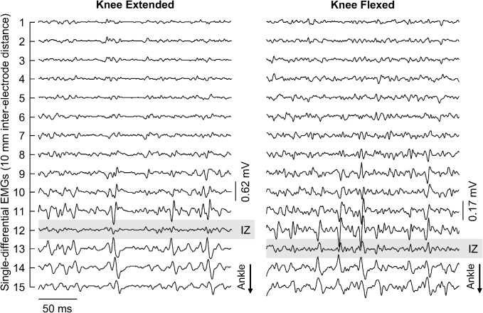
Fig 2. Displacement of innervation zone with knee flexion.
Short epochs (250 ms) of the 15 single-differential EMGs collected from a single participant are shown. Signals in the left and right panels were obtained during knee extended and knee flexed positions, respectively. Propagating potentials are observed in the most distal channels, which were covering the most distal MG fibres. The channel in the array positioned most closely to the innervation zone of the muscle distal fibres is indicated with grey, shaded rectangles. Note the innervation zone moved distally from knee extended to knee flexed position.

